|
||
|
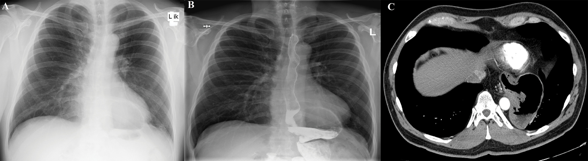
A. The chest X-ray examination in posteroanterior view revealing the presence of hiatal hernia, measuring 95×60 mm, seen as the brightening region which blurred the contour of the diaphragm; B. The chest radiograph with the barium swallow test in the posteroanterior view; C. Computed tomography of the chest in the axial projection revealing the hiatal hernia, measuring 77×90×59 mm with the significant part of the stomach in the thoracic cavity. |