|
||
|
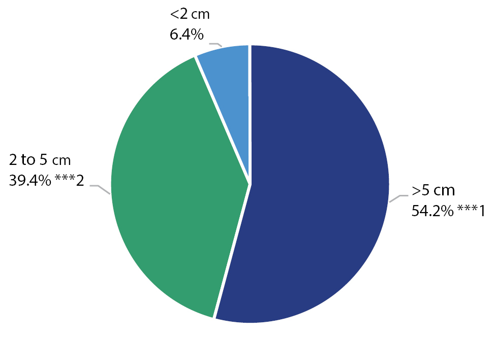
Distribution by size of the peripherally located formation in the chest. ***1 – Significantly higher relative share compared to other sizes (p=0.001); ***2 – Significantly higher relative proportion to tumors smaller than 2 cm (p<0.001). |
|
||||||||
| Part of: Argirov D, Yavorov B, Aleksiev V, Chapkunov A, Shterev F, Kartev S, Uchikov P, Vazhev Z (2024) Complications due to ultrasound transthoracic cutting biopsy of peripheral pulmonary lesions and lesions in the chest wall and mediastinum. Folia Medica 66(2): 179-187. https://doi.org/10.3897/folmed.66.e114030 |