|
||
|
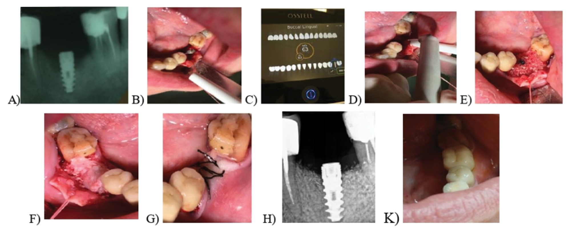
A) Intraoral X-ray of the implant in the area of 36; B) preparation of the flap; C) measured the implant’s stability; D) Granulation tissue ablation and bone remodeling with Er-YAG laser; E) Filling the bone defect with ATB graft material; F) Coating the graft material with PRF membrane; G) adaptation and suturing of the mucoperiosteal flap; H) control radiography after three months; K) Prosthetic rehabilitation of the implant. |