|
||
|
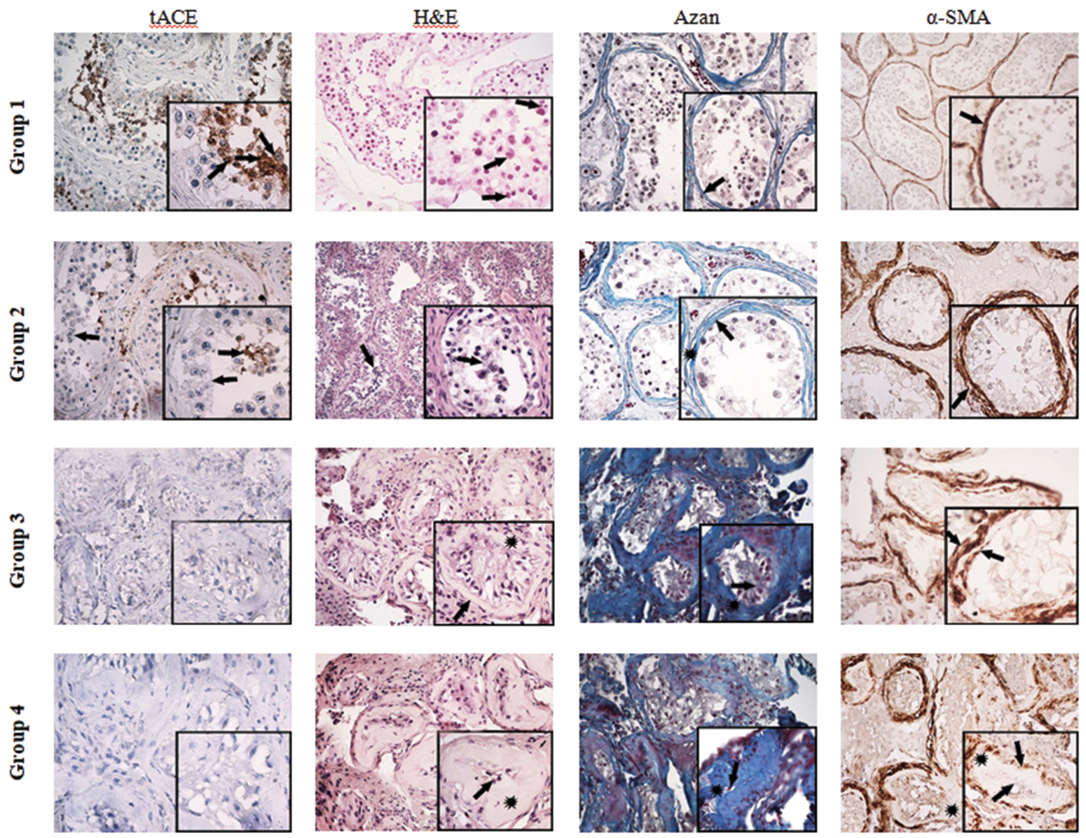
Seminiferous tubules (groups 1-4) – immunoreactivity for tACE and α-SMA, haematoxylin and eosin (H&E), and azan (A) staining; ×100, ×400. Seminiferous tubules of group 1 - (tACE), (H&E), and (A) show normal MP thickness and preserved spermatogenesis; (→) immunoreactivity for α-SMA visualizes myofibroblast cells in MP. By group 2 - reduced tACE expression in individual seminiferous tubules accompanied by disorganization and exfoliation of the spermatogenic epithelium in the lumen (→). (A) and α-SMA show an increase in MP thickness. Lymphocytic infiltration in the testicular interstitium is found. Group 3 - low tACE expression in single round spermatids, (H&E) and (A) outline distinct ES. α-SMA in MP clearly distinguishes two layers of myofibroblasts - outer and inner (→); ECM (*) is deposited between them. There is no specific organization of EI, presence of lymphocyte infiltration. Group 4 - tACE is not visualized; (H&E) and (A) show a reduced lumen with single ES; α-SMA visualizes the absence of an inner layer of myofibroblasts, and only the presence of an outer layer, composed of several myofibroblast layers. |