|
||
|
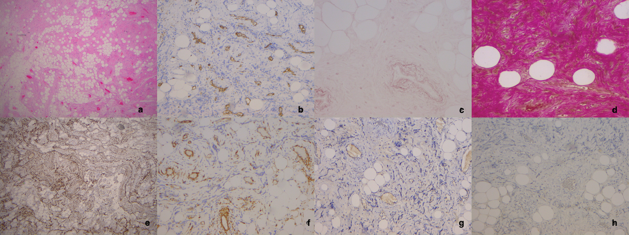
ALLM, although histologically similar to the renal angiomyolipoma, is not associated with tubular sclerosis and does not express melanocytic markers immunohistochemically (HMB-45 and Melan-A). The histological diagnosis is mostly morphological, while the differential diagnosis is based on immunohistochemistry: a) Microscopically we see an indeterminate mixture of smooth muscle tissue, mature adipose tissue and abundant, large, thick-walled, irregular vessels. b) CD34 paints all the vessels but is negative in smooth muscle tissue. It also differentiates from other spindle-shaped neoplasms in which it is positive. c, d) Orcein and Van Gieson stains are histochemical. The Van Gieson stain dyes collagen intensely reddish (also other elements of connective tissue and muscle, but fainter). Both dyes are used in our case to demonstrate the abnormal configuration of collagen within the vessel wall which is characteristic of ALLM. e, f) Smooth muscle tissue markers, desmin (e) more specific than SMA (f), indicate the neoplastic muscle element. g,h) Melanocyte markers (positive in angiomyolipoma-negative here). Their negativity is compatible with the diagnosis of ALLM. |