|
||
|
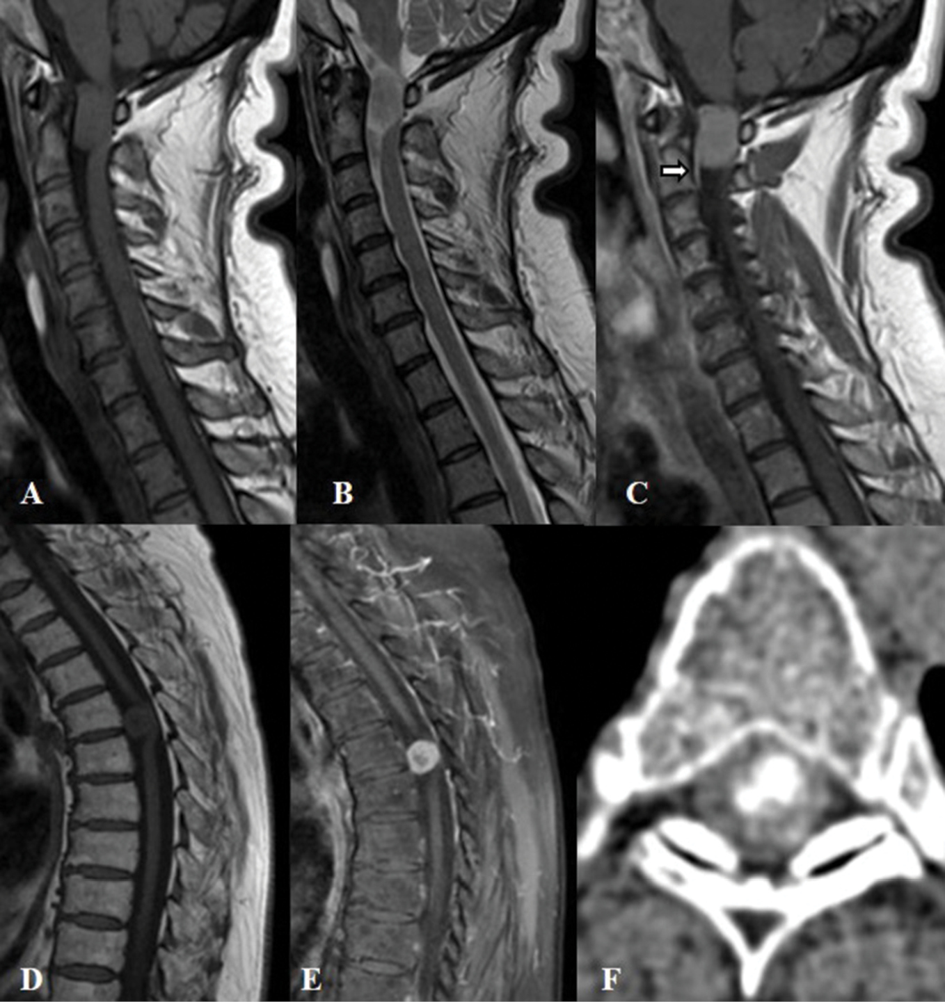
A-C) T1, T2 and enhanced T1 MRI demonstrates antero-laterally located meningioma at the level of C1-C2 with typical “dural tail” sign after gadolinium administration (arrow); D and E) T1 MRI before and after gadolinium administration demonstrates meningioma at the level of T3. On T1 MRI, the tumor is isointense with a hypointense center. The contrast media is accumulated at the periphery of the lesion, but not in the center, due to the presence of calcification; F) Axial CT of the same patient showing the presence of calcification. |