|
||
|
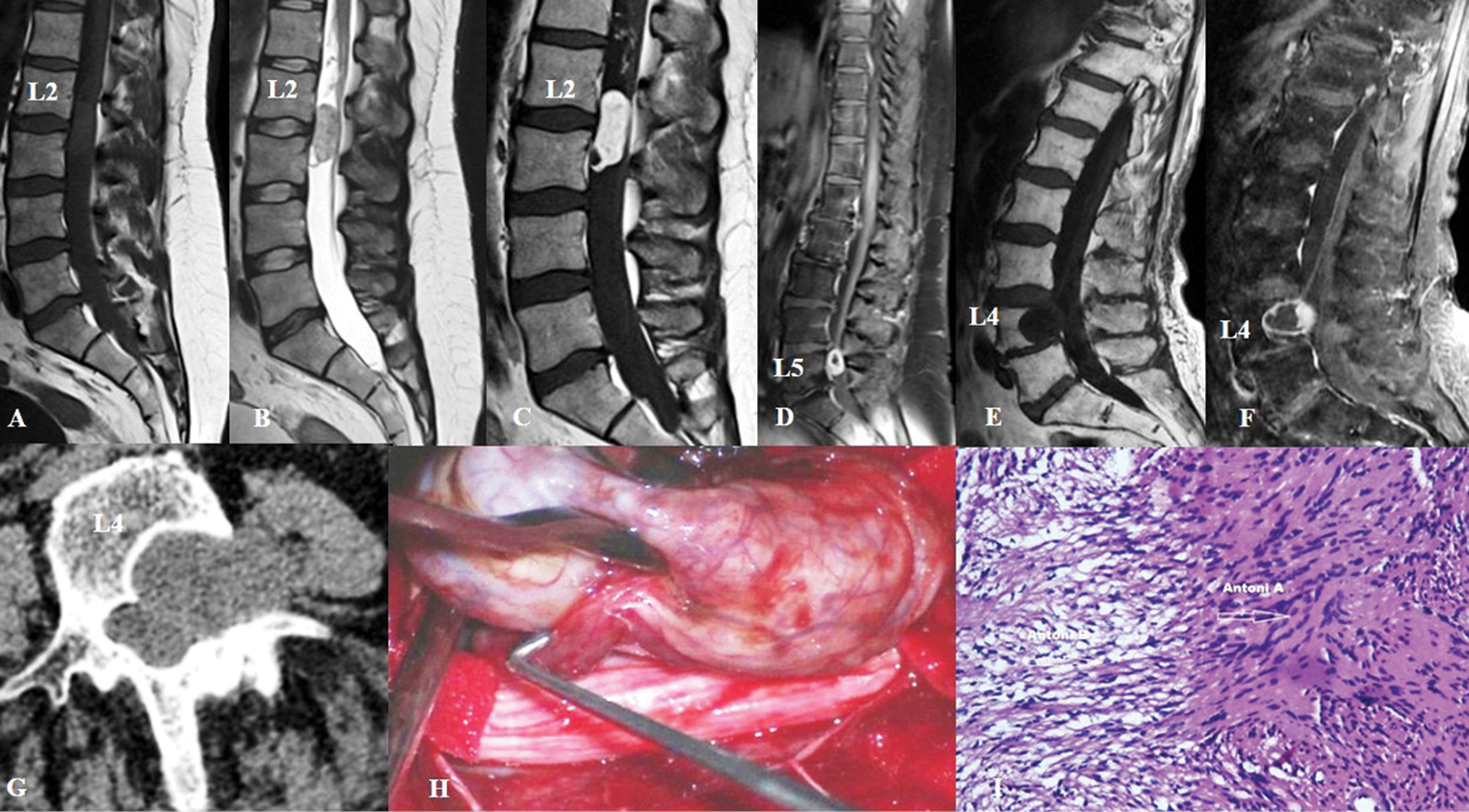
A-C) T1, T2 MRI and enhanced T1 MRI of schwannoma at the level of L2–L3 demonstrating hypointense signal on T1, moderately hyperintense signal on T2, and after contrast - pronounced non-homogeneous accumulation; D) T1 MRI with contrast accumulated at the periphery of the lesion; E and F) T1 MRI before and after enhancement which demonstrates neurofibroma at the level of L4 – non-homogeneous signal amplification of solid components with central cystic region; G) Axial CT of an dumbbell-type neurofibroma at the level of L4 shows enlargement of the left neuroforamen by the tumor and its propagation to the psoas major muscle; H) Intraoperative image; I) Histological specimen consistent with schwannoma; Hematoxylin-Eosin staining, magnification ×100. |