|
||
|
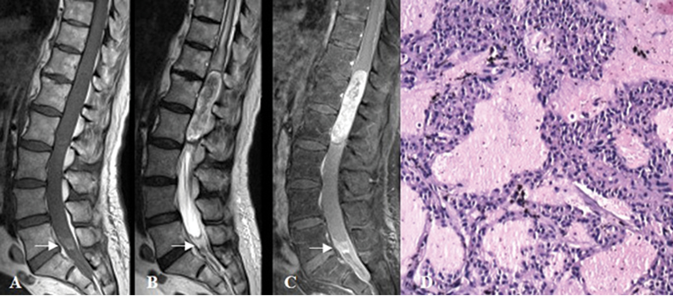
MRI of myxopapillary ependymoma (Grade II) at the level of T12-L2, with drop metastasis (arrows) visualized at the level of L5–S1. A) T1 MRI shows an isointense lesion; B) T2 MRI - the lesion is heterointense and extends downward to conus medullaris; C) Enhanced T1 MRI demonstrates heterogeneous accumulation of the contrast media; D) Histological specimen consistent with myxopapillary ependymoma; Hematoxylin-Eosin staining, magnification ×100. |