|
||
|
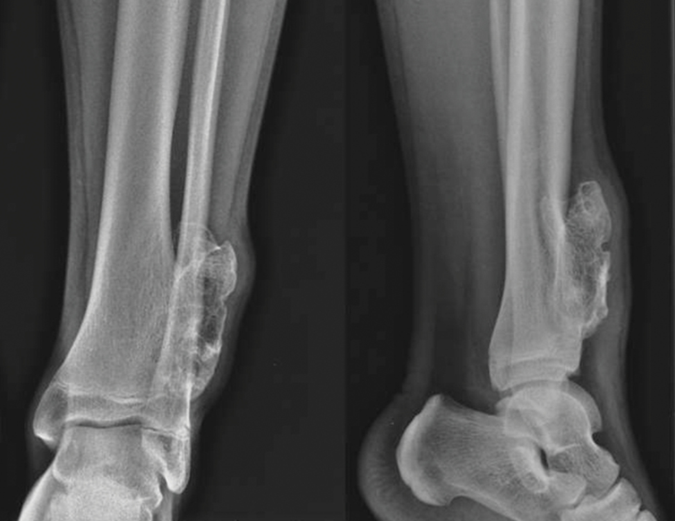
Anteroposterior and lateral radiographs demonstrated a well-defined bone lesion with a broad area of osseous continuity with the parent bone, protruding from the anterolateral distal tibial metaphysis and growing away from the ankle joint. The tumour was composed of cortical and medullary bone, which was in continuity with the tibial marrow cavity. |