|
||
|
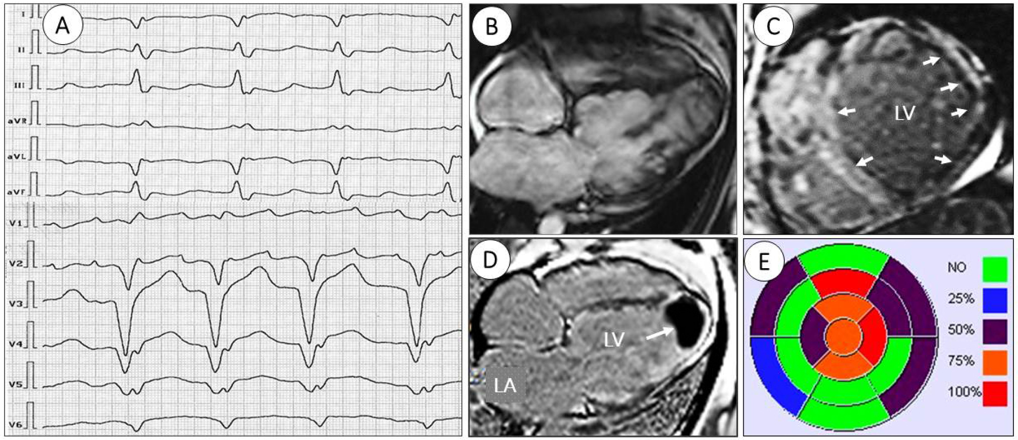
Cardiac anomalies identified in the proband. А. Electrocardiogram of the proband demonstrating atrial flutter, atypical left bundle branch block with pseudo-infarction signs of Sodi-Pollares (abnormal QS in leads I, aVL, V5-V6) and Cabrera sign (notch on the ascending S wave in lead V4 with a duration of 40 ms); B. Cardiac MRI plan the 4-chamber cine on the long axis image shows aneurysmal bulging of left ventricular apex with thrombus; C. T1-native mapping with signs of apical aneurysm and thrombus 34×27 mm; D. Late-gadolinium enhancement imaging on the short axis indicates presence of midwall myocardial contrast delay pattern with extensive linear fibrosis of left ventricular free wall, anterio-inferio-lateralis and septal myocardial scarring (arrowheads); E. Tissue LV characteristic: bull’s eye map image demonstrates late gadolinium enhancement (short axis, 16 segments; grade 0-100%) a diffuse pattern of intramural and transmural fibrosis in the apex, anterior and anterolateral LV wall. |