|
||
|
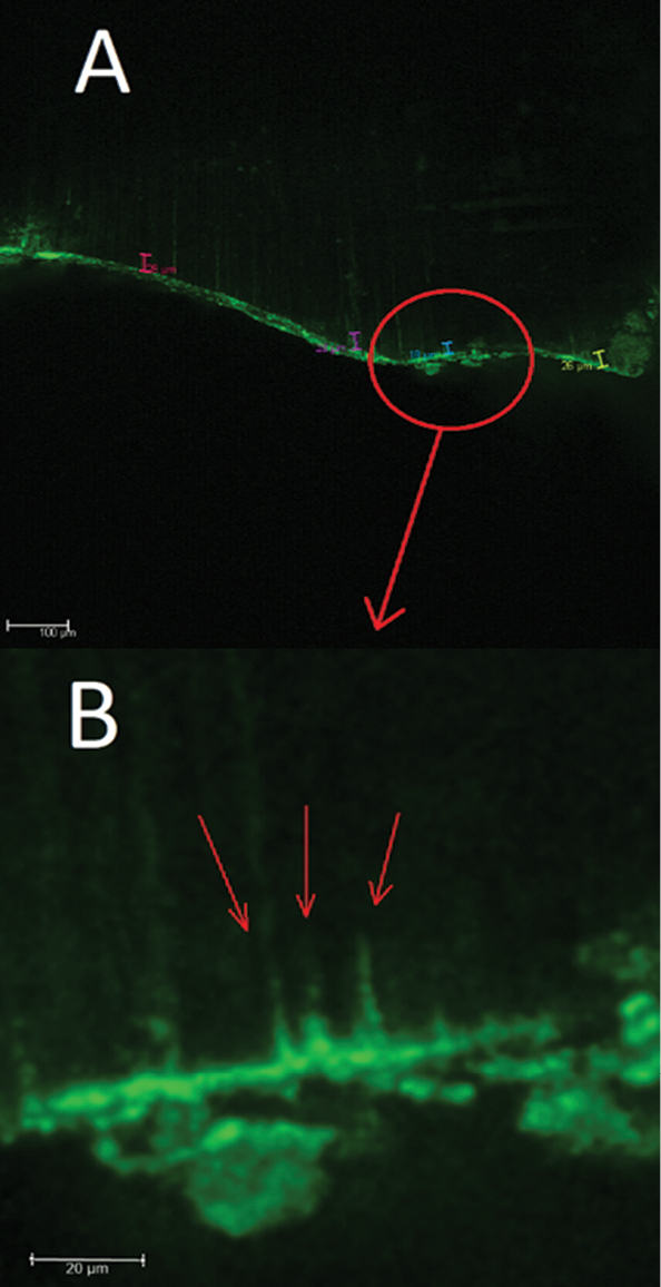
Image of a sample of group 3 (controls). A) Visualization of superimposed image from channel 2 (bond in green) (magnification ×10); B) area further enlarged digitally. Resin-tags are indicated by arrows. |
|
||||||||
| Part of: Simeonov N, Stefanova V (2023) Micromorphology and analysis of dentin surfaces after preparation with Er:YAG laser and application of self-etch adhesive system with 10-MDP. A confocal laser scanning microscope study. Folia Medica 65(3): 453-459. https://doi.org/10.3897/folmed.65.e76606 |