|
||
|
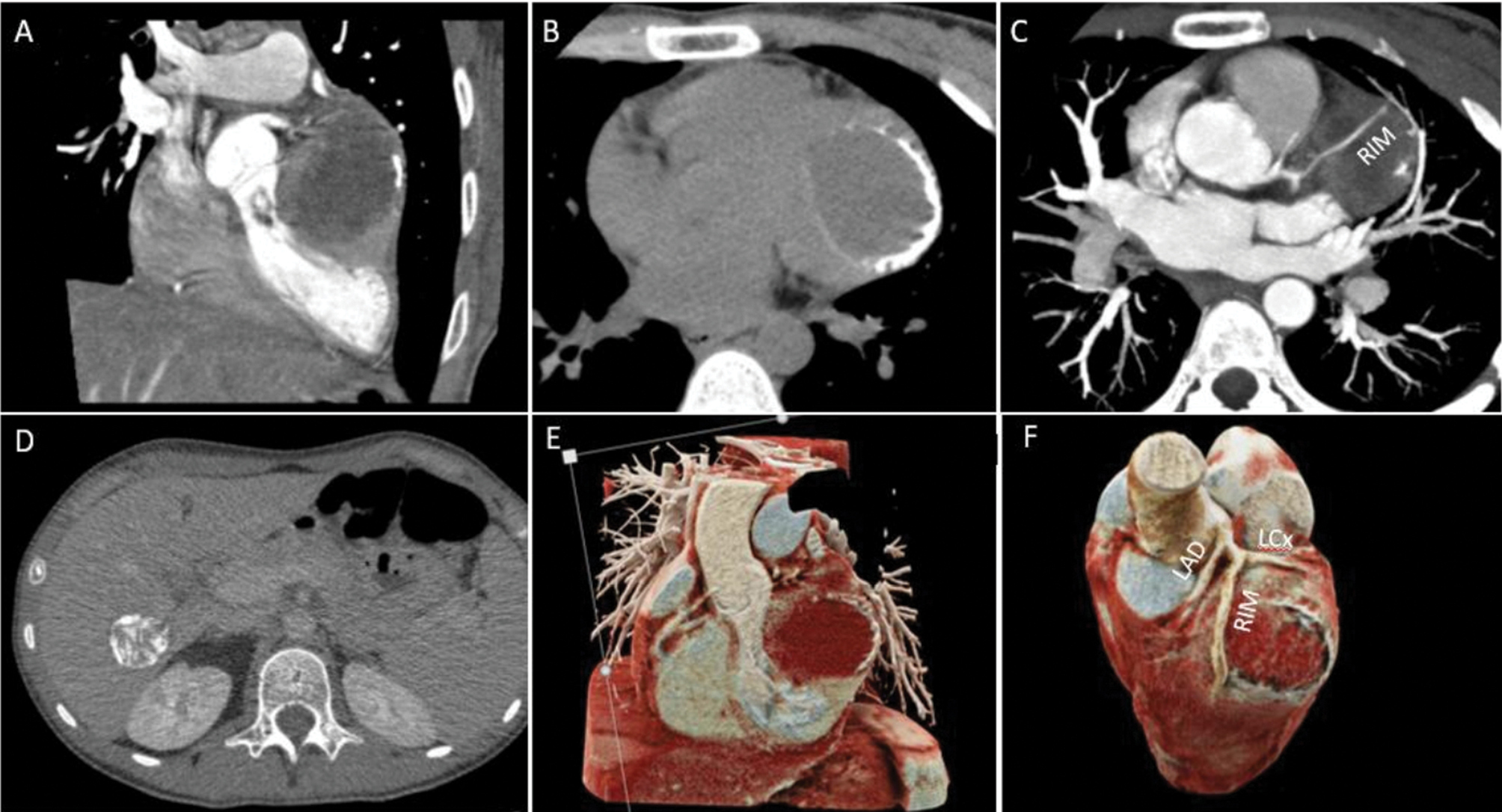
Computed tomography demonstrating the cystic lesion in the heart, located in the left ventricle lateral wall with additional 3D cinematic VRT. The typical peripheral calcifications are well demonstrated (B). RIM passing over the cyst (C). Liver lesion with typical water-lily sign is also shown (D). A coronal view of the cyst (E) and volume rendering of coronary arteries (F). LAD: left anterior descending artery; LCx: left circumflex artery; RIM: ramus intermedius. |