|
||
|
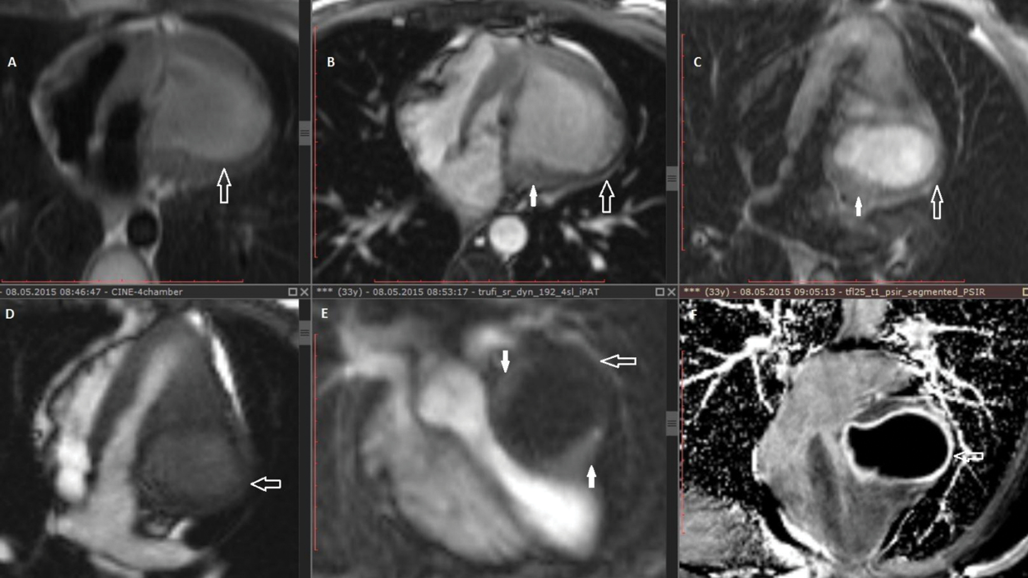
CMR images demonstrating cystic lesion in the lateral free wall of the left ventricle. The cyst shows high signal on HASTE (A), SSFP (B), TIRM (C) sequences and is surrounded by a thin hypointense rim (C). Dynamic first pass myocardial perfusion (D, E) shows no enhancement of the intramyocardial mass and in the same time well depicts the intramyocardial location of the lesion, showing the normally perfused myocardium surrounding the lesion (E - white arrows). Late gadolinium enhancement shows intense enhancement in the fibrous capsule (F). |