|
||
|
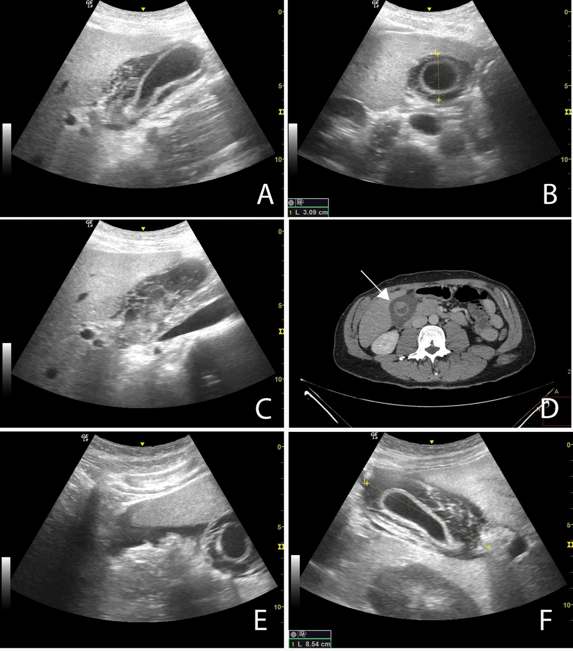
A. Ultrasound image, longitudinal section of the gallbladder; B. Ultrasound image, cross-section of the gallbladder; C. Ultrasound image, imbibition of the pericholecystic tissue; D. CT scan of abdomen, (gallbladder and pericholecystic fat tissue marked with an arrow); E. Ultrasound image, free fluid under the liver; F. Ultrasound image, perivesical hematoma. |